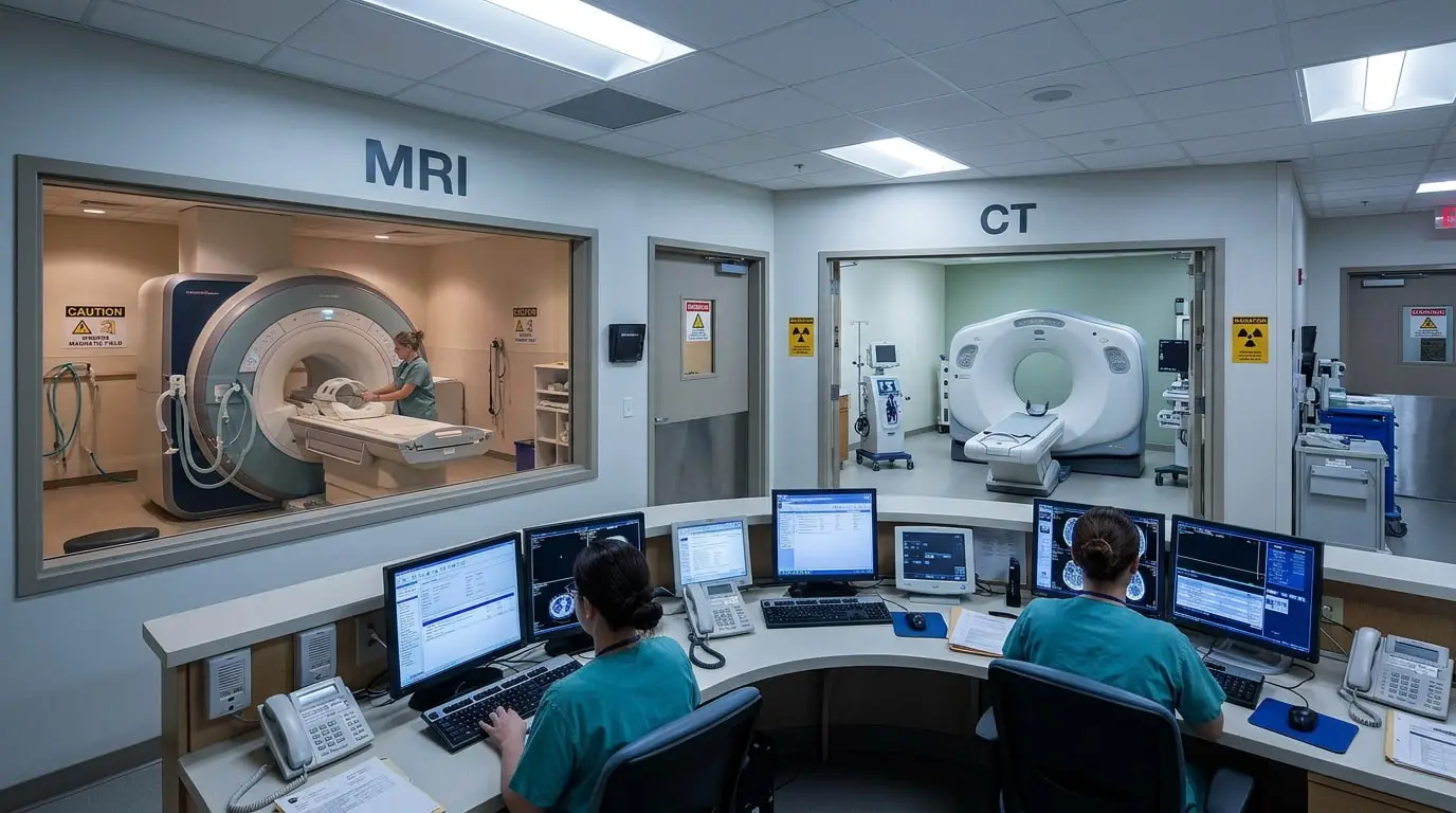
MRI vs CT Scan: Understanding the Right Test for Your Health
When a doctor recommends medical imaging, patients often wonder which test they should undergo and why. Understanding the differences between MRI vs CT scan can help you feel more prepared before your appointment and understand how each examination contributes to diagnosing a medical condition.
Although both imaging techniques provide detailed pictures of internal structures, they use different technologies and are suited to different diagnostic needs. The choice depends on the body area being examined, the suspected condition, and how quickly the doctor needs the results.
At Gardenia Medical Centre in Doha, diagnostic imaging services help support medical evaluations by providing access to different imaging techniques through our specialized Diagnostic and Interventional Radiology Department. Understanding the purpose of each examination can help patients make informed decisions about their diagnostic care in consultation with their doctor.
What Is an MRI Scan and How Does It Work?
Magnetic Resonance Imaging (MRI) is a diagnostic imaging technique that uses a powerful magnetic field and radio waves to produce detailed images of organs, muscles, nerves, and other internal structures.
Unlike X-rays, MRI does not use ionizing radiation. It is particularly valuable when doctors need detailed information about soft tissues, including structures that may not appear clearly on other imaging examinations.
An MRI scan for soft tissue assessment may be recommended to examine:
- The brain, spinal cord, and cranial/peripheral nerves
- Ligaments, tendons, cartilage, and muscles
- Intervertebral discs and intricate spinal structures
- Certain internal organs (liver, pelvis, kidneys)
- Tumors, vascular malformations, and subtle inflammatory changes
MRI is commonly used to investigate neurological conditions, joint injuries, spinal problems, and certain abnormalities involving internal organs. However, patients with implanted medical devices (such as pacemakers or cochlear implants) or metallic foreign objects must undergo appropriate safety screening before entering the MRI room.[1]
For more information about available examinations, visit our MRI Scan Centre in Qatar page.
What Is a CT Scan and How Does It Work?
Computed Tomography (CT) uses X-rays taken from multiple angles to create cross-sectional images of the body. A computer combines these slices to provide detailed views of bones, organs, blood vessels, and other internal structures.
CT examinations are particularly useful when a doctor needs detailed images quickly, especially in emergency situations. A CT scan for bone fractures can help evaluate complex injuries, identify the extent of a fracture, and provide 3D reconstructions that support surgical planning.
Doctors may also recommend CT imaging for:
- Head injuries, suspected intracranial hemorrhage, and acute strokes
- Chest and lung conditions (pulmonary embolism, pneumonia, nodules)
- Acute abdominal and pelvic problems (appendicitis, diverticulitis, bowel obstruction)
- Complex bone fractures and joint trauma
- Tumor staging, response monitoring, and follow-up
- Internal trauma and vascular injuries following accidents
One of the main advantages of CT is its remarkable speed. However, because it uses ionizing radiation, doctors consider the medical necessity of the examination and aim to use an appropriate, optimized radiation dose under the ALARA (As Low As Reasonably Achievable) principle.[2]
You can learn more about the examination through our CT Scan in Qatar page.
MRI vs CT Scan: What Are the Main Differences?
The main differences between these imaging techniques involve their physical technology, the tissue structures they depict with greatest clarity, examination duration, and radiation safety considerations:
| Feature | MRI Scan | CT Scan |
|---|---|---|
| Imaging Technology | Powerful magnetic fields and radiofrequency waves | Multiple rotating X-ray beams and computer detectors |
| Ionizing Radiation | No (Completely radiation-free) | Yes (Low-dose ionizing radiation) |
| Soft Tissue Imaging | Superior soft tissue contrast and anatomical detail | Good general detail, but less tissue differentiation |
| Bone Imaging | Excellent for bone marrow edema and surrounding cartilage | Outstanding for cortical bone, acute fractures, and calcifications |
| Examination Time | Longer (typically 15 to 45 minutes) | Very fast (typically 2 to 10 minutes) |
| Emergency Suitability | Limited in trauma; reserved for specific neurological issues | Primary gold standard for acute emergency and trauma imaging |
| Contrast Material | Gadolinium-based contrast agents | Iodine-based contrast agents |
Understanding these differences helps explain why doctors may recommend one examination instead of the other depending on the diagnostic target.[1]
Which Is More Accurate: MRI or CT?
There is no single answer to whether MRI or CT is more accurate. Each technique provides different diagnostic information, and its clinical value depends entirely on the tissue or pathology being investigated.
MRI generally provides superior contrast resolution for soft tissues, making it particularly useful for evaluating ligament tears, meniscal damage, spinal disc herniations, and delicate neurological conditions. CT is often significantly more effective for identifying complex fractures, evaluating acute chest trauma, and detecting fresh internal bleeding when rapid intervention is needed.
For example, an MRI is ideal for a suspected knee ligament injury (such as an ACL tear), while a CT scan provides far better diagnostic precision regarding the bone fragment alignment of a complicated tibia fracture. Therefore, the most appropriate examination is the one that directly answers the clinician's specific diagnostic question.[2]
When Does a Doctor Recommend CT Instead of MRI?
Doctors typically order a CT scan when they need rapid acquisition or detailed evaluation of cortical bones, vascular patency, and acute traumatic injuries:
- Head injuries: CT rapidly detects intracranial hemorrhage, hematomas, and skull fractures.
- Complex fractures: CT provides high-resolution 3D bone mapping.
- Acute abdominal pain: Quickly rules in or out appendicitis, kidney stones, or bowel obstruction.
- Chest conditions: Evaluates lung parenchyma, interstitial disease, and pulmonary embolism.
- Internal bleeding: Quickly isolates hemorrhagic sources in polytrauma patients.
When Is MRI the Better Choice?
MRI is the modality of choice when soft tissues, the central nervous system, joints, or pelvic organs require meticulous characterization:
- Brain lesions, multiple sclerosis plaques, and complex neurological diseases
- Spinal cord compression, herniated intervertebral discs, and nerve root impingement
- Sports-related ligament tears, tendon ruptures, and articular cartilage lesions
- Deep muscle tears, soft tissue sarcomas, and muscular inflammation
- Detailed evaluation of pelvic anatomy (uterine fibroids, prostate staging, endometriosis)
- Tumours and surrounding tissue changes
Contrast Agents, Safety Profiles, and Pregnancy Guidelines
MRI With Contrast vs CT With Contrast
Contrast agents improve the diagnostic conspicuity of vascularity, tissue boundaries, and inflammatory lesions:
- MRI with contrast: Typically utilizes gadolinium-based agents administered intravenously. Gadolinium alters the relaxation times of adjacent water protons, highlighting areas of hypervascularity, active breakdown of the blood-brain barrier, or active inflammation.
- CT with contrast: Generally utilizes iodinated contrast media that attenuate X-rays, making blood vessels (CT angiography), visceral perfusion, and solid organ lesions radiopaque and clearly visible.
Not every scan requires contrast. Prior to contrast administration, patients must disclose history of renal impairment, known drug allergies, or prior reactions to contrast media.[3]
Is MRI Safer Than CT?
Both examinations are routine and clinically safe, but their risk profiles differ fundamentally. Because MRI does not use ionizing radiation, it avoids radiation-associated risks entirely. However, the powerful static magnetic field presents strict physical safety constraints: any ferromagnetic implant, pacemaker, or metallic fragment could be displaced or cause thermal injury, requiring rigorous pre-scan safety screening.
CT scans involve a controlled dose of ionizing radiation. While modern multidetector scanners employ iterative reconstruction algorithms to minimize exposure, scans should still be clinically justified. Iodinated CT contrast also carries a slightly higher risk of allergic reactions and nephrotoxicity compared to gadolinium agents.[2]
Pregnancy Considerations: MRI without contrast is generally favored during pregnancy when advanced cross-sectional imaging is required, as it avoids fetal radiation exposure. CT is not contraindicated if an acute maternal emergency (e.g., pulmonary embolism, severe trauma) demands rapid diagnosis. Gadolinium contrast is avoided during pregnancy due to potential fetal risks unless clinically essential.[4]
How to Prepare for an MRI or CT Scan and Receiving Results
Preparation Guidelines
Preparation depends on the anatomical region examined and whether contrast administration is indicated:
- Before an MRI: Complete the metal and implant safety screening form. Remove all jewelry, body piercings, watches, hairpins, and metallic clothing. Notify the technologist of claustrophobia, pregnancy, or medical implants. Follow fasting protocols if abdominal/pelvic imaging is scheduled.
- Before a CT Scan: Inform the radiology team of any prior allergic reactions to iodinated contrast or shellfish. Fast for 2 to 4 hours if intravenous contrast is ordered. Disclose current medications, particularly metformin if kidney disease is present.
Never discontinue prescribed maintenance medication without direct physician instruction.[5]
When Will Results Be Available?
Although a CT acquisition takes only minutes and an MRI may take half an hour, the timing of the final report depends on careful clinical review. A specialist radiologist must thoroughly analyze the multi-planar image series, compare them against previous studies, and issue a formal diagnostic report to your referring physician.
Diagnostic and Interventional Radiology at Gardenia Medical Centre
Gardenia Medical Centre provides comprehensive diagnostic imaging in Doha, combining cutting-edge scanner technology with high-caliber clinical interpretation. Under the direction of experienced diagnostic and interventional radiologists, our imaging services support precise diagnosis across orthopedics, neurology, internal medicine, and surgery.

Dr. Abdul Khalek Mohamed El Ghol
Consultant in Diagnostic and Interventional Radiology | Head of Department
Dr. Abdul Khalek Mohamed El Ghol heads the Diagnostic and Interventional Radiology Department. He brings extensive clinical expertise in cross-sectional diagnostic imaging (CT and MRI), body imaging, and minimally invasive image-guided interventions.
His dedicated approach ensures that every imaging procedure is selected appropriately to answer the clinical question while adhering to the highest international radiation and patient safety protocols.
Beyond advanced cross-sectional CT and MRI examinations, our radiology department offers integrated diagnostic support, including digital X-rays, Doppler vascular studies, diagnostic ultrasound, and specialized musculoskeletal assessments.
Book Your Diagnostic Imaging Appointment in Doha
If your doctor has recommended an MRI or CT examination, early scheduling and proper preparation ensure an accurate and seamless diagnostic journey. Contact Gardenia Medical Centre today to schedule your examination:
- Explore our full range of Diagnostic and Interventional Radiology Services.
- Get in touch directly through our Contact Page.
- Schedule your consultation via the Online Appointment Booking Page.
Frequently Asked Questions About MRI vs CT Scan
Are MRI and CT scans the same in Qatar?
No. MRI uses a magnetic field and radiofrequency waves without radiation, whereas a CT scan uses rotating X-rays and detectors. Each modality serves distinct clinical and diagnostic purposes.
Which is better for brain imaging in Doha, MRI or CT?
CT is the primary choice for acute trauma, suspected hemorrhage, or emergency stroke triage due to its speed. MRI is superior for non-emergency brain assessment, subtle ischemia, tumors, multiple sclerosis, and nerve root evaluation.
Does a CT scan in Qatar always require contrast?
No. Many CT scans—such as those for kidney stones, non-contrast head trauma, or bone fractures—do not require contrast. Contrast is requested when evaluating blood vessels, soft tissue perfusion, or oncologic staging.
Can MRI detect tumours in Doha?
Yes. MRI provides exquisite contrast resolution that allows radiologists to detect tumors, define their precise margins, and evaluate involvement with adjacent vascular or neurological structures.
Can MRI be performed after a CT scan in Qatar?
Yes. Doctors frequently order an MRI following an initial CT scan when complementary soft-tissue detail, spinal cord imaging, or clearer characterization of an ambiguous finding is required.
References
- FDA - Benefits and Risks of Magnetic Resonance Imaging (MRI)
- FDA - Computed Tomography (CT) Overview and Radiation Safety
- RadiologyInfo.org - Contrast Materials Overview, Indications & Safety
- ACOG - Guidelines for Diagnostic Imaging During Pregnancy and Lactation
- RadiologyInfo.org - Patient Preparation Guidelines for MRI Examinations